Regenerative endodontics
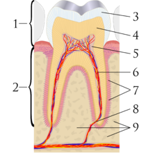
Cross section of a tooth: (1)crown (2)root (3)enamel (4)denting & dentin tubules (5)pulp chamber (6)blood vessels & nerve (7)periodontal ligament (8)apex (9)alveolar bone
Regenerative endodontics is the use of biologically based procedures designed to replace damaged tooth structures such as dentin, root structures and cells of the pulp-dentin complete.[1] Regenerative endodontics is the extension of root canal therapy. Conventional root canal therapy cleans and fills the pulp chamber with biologically inert material after destruction of the pulp due to dental caries, congenital deformity or trauma. Regenerative endodontics instead seeks to replace live tissue in the pulp chamber.
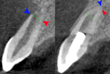
Regenerative endodontics in 10-year-old with necrotic pulp and incomplete root formation (left) then 1 year after treatment. Buccal aspect of apex (blue arrow), palatal aspect of apex (red arrow) and line of initial root formation (green line)
To replace live tissue, either the existing cells of the body are stimulated to regrow the tissue native to the area or bioactive substances inserted in the pulp chamber. These include stem cell therapy, growth factors, morphogens, tissue scaffolds and biologically active delivery systems.[2]
Closely related to the field of regenerative endodontics, are the clinical procedures apexification and apexogenesis. When the dental pulp of a developing adult tooth dies, root formation is halted leaving an open tooth apex. Attempting to complete root canal on a tooth with an open apex is technically difficult and the long-term prognosis for the tooth is poor.
Apexogenesis, (which can be used when the pulp is injured but not necrotic) leaves the apical one-third of the dental pulp in the tooth which allows the root to complete formation. Apexification, stimulates cells in the periapical area of the tooth to form a dentin-like substance over the apex. Both improve the long-term prognosis for a forming tooth over root canal alone.[3]
Necrotic pulp and open apex can be revitalized with platelet rich fibrin.[4]

