Difference between revisions of "Reactive Oxygen Species(ROS)"
imported>Kyunghyun Cho (Created page with "<h1>Reactive oxygen species</h1> <p>From Wikipedia, the free encyclopedia</p> <p><a href="https://en.wikipedia.org/wiki/File:Major_cellular_sources_of_Reactive_Oxygen_Species_i...") |
(No difference)
|
Revision as of 17:14, 1 December 2018
Contents
- 1 Reactive oxygen species
- 1.1 -----------------------------------------------------------------------------------------------------------------------------------------------------------------------
- 1.2 Formation and decomposition
- 1.3 -----------------------------------------------------------------------------------------------------------------------------------------------------------------------
- 1.4 Damaging effects
Reactive oxygen species
From Wikipedia, the free encyclopedia
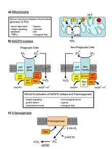
Major cellular sources of ROS in living non-photosynthetic cells. From a review by Novo and Parola, 2008.[1]
Reactive oxygen species (ROS) are chemically reactive chemical species containing oxygen. Examples include peroxides, superoxide, hydroxyl radical, singlet oxygen,[2] and alpha-oxygen.
In a biological context, ROS are formed as a natural byproduct of the normal metabolism of oxygen and have important roles in cell signaling and homeostasis.[3] However, during times of environmental stress (e.g., UV or heat exposure), ROS levels can increase dramatically.[3] This may result in significant damage to cell structures. Cumulatively, this is known as oxidative stress. The production of ROS is strongly influenced by stress factor responses in plants, these factors that increase ROS production include, drought, salinity, chilling, nutrient deficiency, metal toxicity and UV-B radiation. ROS are also generated by exogenous sources such as ionizing radiation.[4]
-----------------------------------------------------------------------------------------------------------------------------------------------------------------------
Formation and decomposition
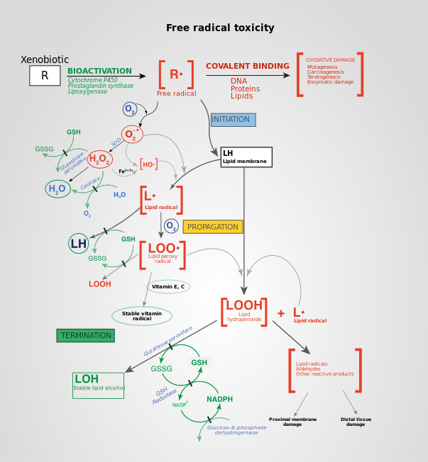
Free Radical Mechanisms in Tissue Injury. Free radical toxicity induced by xenobiotics and the subsequent detoxification by cellular enzymes (termination).
The reduction of molecular oxygen (O2) produces superoxide (•O−2) and is the precursor of most other reactive oxygen species:[5]
O2 + e− → •O−2
Dismutation of superoxide produces hydrogen peroxide (H2O2):[5]
2 H+ + •O−
2 + •O−
2 → H2O2 + O2
Hydrogen peroxide in turn may be partially reduced to hydroxyl radical (•OH) or fully reduced to water:[5]
H2O2 + e− → HO− + •OH
2 H+ + 2 e− + H2O2 → 2 H2O
Exogenous ROS
Exogenous ROS can be produced from pollutants, tobacco, smoke, drugs, xenobiotics, or radiation.
Ionizing radiation can generate damaging intermediates through the interaction with water, a process termed radiolysis. Since water comprises 55–60% of the human body, the probability of radiolysis is quite high under the presence of ionizing radiation. In the process, water loses an electron and becomes highly reactive. Then through a three-step chain reaction, water is sequentially converted to hydroxyl radical (•OH), hydrogen peroxide (H2O2), superoxide radical (•O−2), and ultimately oxygen (O2).
The hydroxyl radical is extremely reactive and immediately removes electrons from any molecule in its path, turning that molecule into a free radical and thus propagating a chain reaction. However, hydrogen peroxide is actually more damaging to DNA than the hydroxyl radical, since the lower reactivity of hydrogen peroxide provides enough time for the molecule to travel into the nucleus of the cell, subsequently reacting with macromolecules such as DNA.
Endogenous ROS
ROS are produced intracellularly through multiple mechanisms and depending on the cell and tissue types, the major sources being the "professional" producers of ROS: NADPH oxidase (NOX) complexes (7 distinct isoforms) in cell membranes, mitochondria, peroxisomes, and endoplasmic reticulum.[6][7] Mitochondria convert energy for the cell into a usable form, adenosine triphosphate (ATP). The process in which ATP is produced, called oxidative phosphorylation, involves the transport of protons (hydrogen ions) across the inner mitochondrial membrane by means of the electron transport chain. In the electron transport chain, electrons are passed through a series of proteins via oxidation-reduction reactions, with each acceptor protein along the chain having a greater reduction potential than the previous. The last destination for an electron along this chain is an oxygen molecule. In normal conditions, the oxygen is reduced to produce water; however, in about 0.1–2% of electrons passing through the chain (this number derives from studies in isolated mitochondria, though the exact rate in live organisms is yet to be fully agreed upon), oxygen is instead prematurely and incompletely reduced to give the superoxide radical (•O−2), most well documented for Complex I and Complex III.[8] Superoxide is not particularly reactive by itself, but can inactivate specific enzymes or initiate lipid peroxidation in its protonated form, hydroperoxyl HO•2. The pKa of hydroperoxyl is 4.8. Thus, at physiological pH, the majority will exist as superoxide anion.
If too much damage is present in mitochondria, a cell undergoes apoptosis or programmed cell death.[9][10] Bcl-2 proteins are layered on the surface of the mitochondria, detect damage, and activate a class of proteins called Bax, which punch holes in the mitochondrial membrane, causing cytochrome C to leak out. This cytochrome C binds to Apaf-1, or apoptotic protease activating factor-1, which is free-floating in the cell's cytoplasm. Using energy from the ATPs in the mitochondrion, the Apaf-1 and cytochrome C bind together to form apoptosomes. The apoptosomes bind to and activate caspase-9, another free-floating protein. The caspase-9 then cleaves the proteins of the mitochondrial membrane, causing it to break down and start a chain reaction of protein denaturation and eventually phagocytosis of the cell.
Superoxide dismutase
Main article: Superoxide dismutase
Superoxide dismutases (SOD) are a class of enzymes that catalyze the dismutation of superoxide into oxygen and hydrogen peroxide. As such, they are an important antioxidant defense in nearly all cells exposed to oxygen. In mammals and most chordates, three forms of superoxide dismutase are present. SOD1 is located primarily in the cytoplasm, SOD2 in the mitochondria and SOD3 is extracellular. The first is a dimer (consists of two units), while the others are tetramers (four subunits). SOD1 and SOD3 contain copper and zinc ions, while SOD2 has a manganese ion in its reactive centre. The genes are located on chromosomes 21, 6, and 4, respectively (21q22.1, 6q25.3 and 4p15.3-p15.1).
The SOD-catalysed dismutation of superoxide may be written with the following half-reactions:
- M(n+1)+ − SOD + O−2 → Mn+ − SOD + O2
- Mn+ − SOD + O−2 + 2H+ → M(n+1)+ − SOD + H2O2.
where M = Cu (n = 1); Mn (n = 2); Fe (n = 2); Ni (n = 2). In this reaction the oxidation state of the metal cation oscillates between n and n + 1.
Catalase, which is concentrated in peroxisomes located next to mitochondria, reacts with the hydrogen peroxide to catalyze the formation of water and oxygen. Glutathione peroxidasereduces hydrogen peroxide by transferring the energy of the reactive peroxides to a very small sulfur-containing protein called glutathione. The sulfur contained in these enzymes acts as the reactive center, carrying reactive electrons from the peroxide to the glutathione. Peroxiredoxins also degrade H2O2, within the mitochondria, cytosol, and nucleus.
- 2 H2O2 → 2 H2O + O2 (catalase)
- 2GSH + H2O2 → GS–SG + 2H2O (glutathione peroxidase)
Singlet oxygen
Another type of reactive oxygen species is singlet oxygen (1O2) which is produced for example as byproduct of photosynthesis in plants. In the presence of light and oxygen, photosensitizers such as chlorophyll may convert triplet (3O2) to singlet oxygen:[11]
{\displaystyle {\ce {^3O2 ->[{\ce {light}}][{\ce {photosensitizer}}] ^1O2}}}
Singlet oxygen is highly reactive, especially with organic compounds that contain double bonds. The resulting damage caused by singlet oxygen reduces the photosynthetic efficiency of chloroplasts. In plants exposed to excess light, the increased production of singlet oxygen can result in cell death.[11] Various substances such as carotenoids, tocopherols and plastoquinones contained in chloroplasts quench singlet oxygen and protect against its toxic effects. In addition to direct toxicity, singlet oxygen acts a signaling molecule.[11] Oxidized products of β-carotene arising from the presence of singlet oxygen act as second messengers that can either protect against singlet oxygen induced toxicity or initiate programmed cell death. Levels of jasmonate play a key role in the decision between cell acclimation or cell death in response to elevated levels of this reactive oxygen species.[11]
-----------------------------------------------------------------------------------------------------------------------------------------------------------------------
Damaging effects
Effects of ROS on cell metabolism are well documented in a variety of species. These include not only roles in apoptosis (programmed cell death) but also positive effects such as the induction of host defence[12][13]genes and mobilization of ion transport systems.[citation needed] This implicates them in control of cellular function. In particular, platelets involved in woundrepair and blood homeostasis release ROS to recruit additional platelets to sites of injury. These also provide a link to the adaptive immune system via the recruitment of leukocytes.[citation needed]
Reactive oxygen species are implicated in cellular activity to a variety of inflammatory responses including cardiovascular disease. They may also be involved in hearing impairment via cochlear damage induced by elevated sound levels, in ototoxicity of drugs such as cisplatin, and in congenital deafness in both animals and humans.[citation needed] ROS are also implicated in mediation of apoptosis or programmed cell death and ischaemic injury. Specific examples include stroke and heart attack.[citation needed]
In general, harmful effects of reactive oxygen species on the cell are most often:[14]
- damage of DNA or RNA
- oxidations of polyunsaturated fatty acids in lipids (lipid peroxidation)
- oxidations of amino acids in proteins
- oxidative deactivation of specific enzymes by oxidation of co-factors
Pathogen response
When a plant recognizes an attacking pathogen, one of the first induced reactions is to rapidly produce superoxide (O−2) or hydrogen peroxide (H2O2) to strengthen the cell wall. This prevents the spread of the pathogen to other parts of the plant, essentially forming a net around the pathogen to restrict movement and reproduction.
In the mammalian host, ROS is induced as an antimicrobial defense. To highlight the importance of this defense, individuals with chronic granulomatous disease who have deficiencies in generating ROS, are highly susceptible to infection by a broad range of microbes including Salmonella enterica, Staphylococcus aureus, Serratia marcescens, and Aspergillus spp.
The exact manner in which ROS defends the host from invading microbe is not fully understood. One of the more likely modes of defense is damage to microbial DNA. Studies using Salmonella demonstrated that DNA repair mechanisms were required to resist killing by ROS. More recently, a role for ROS in antiviral defense mechanisms has been demonstrated via Rig-like helicase-1 and mitochondrial antiviral signaling protein. Increased levels of ROS potentiate signaling through this mitochondria-associated antiviral receptor to activate interferon regulatory factor (IRF)-3, IRF-7, and nuclear factor kappa B (NF-κB), resulting in an antiviral state.[15] Respiratory epithelial cells were recently demonstrated to induce mitrochondrial ROS in response to influenza infection. This induction of ROS led to the induction of type III interferon and the induction of an antiviral state, limiting viral replication.[16] In host defense against mycobacteria, ROS play a role, although direct killing is likely not the key mechanism; rather, ROS likely affect ROS-dependent signalling controls, such as cytokine production, autophagy, and granuloma formation.[17]
Reactive oxygen species are also implicated in activation, anergy and apoptosis of T cells.[18]
Oxidative damage
In aerobic organisms the energy needed to fuel biological functions is produced in the mitochondria via the electron transport chain. In addition to energy, reactive oxygen species (ROS) with the potential to cause cellular damage are produced. ROS can damage lipid, DNA, RNA, and proteins, which, in theory, contributes to the physiology of aging.
ROS are produced as a normal product of cellular metabolism. In particular, one major contributor to oxidative damage is hydrogen peroxide (H2O2), which is converted from superoxidethat leaks from the mitochondria. Catalase and superoxide dismutase ameliorate the damaging effects of hydrogen peroxide and superoxide, respectively, by converting these compounds into oxygen and hydrogen peroxide (which is later converted to water), resulting in the production of benign molecules. However, this conversion is not 100% efficient, and residual peroxides persist in the cell. While ROS are produced as a product of normal cellular functioning, excessive amounts can cause deleterious effects.[19] Memory capabilities decline with age, evident in human degenerative diseases such as Alzheimer's disease, which is accompanied by an accumulation of oxidative damage. Current studies demonstrate that the accumulation of ROS can decrease an organism's fitness because oxidative damage is a contributor to senescence. In particular, the accumulation of oxidative damage may lead to cognitive dysfunction, as demonstrated in a study in which old rats were given mitochondrial metabolites and then given cognitive tests. Results showed that the rats performed better after receiving the metabolites, suggesting that the metabolites reduced oxidative damage and improved mitochondrial function.[20] Accumulating oxidative damage can then affect the efficiency of mitochondria and further increase the rate of ROS production.[21] The accumulation of oxidative damage and its implications for aging depends on the particular tissue type where the damage is occurring. Additional experimental results suggest that oxidative damage is responsible for age-related decline in brain functioning. Older gerbils were found to have higher levels of oxidized protein in comparison to younger gerbils. Treatment of old and young mice with a spin trapping compound caused a decrease in the level of oxidized proteins in older gerbils but did not have an effect on younger gerbils. In addition, older gerbils performed cognitive tasks better during treatment but ceased functional capacity when treatment was discontinued, causing oxidized protein levels to increase. This led researchers to conclude that oxidation of cellular proteins is potentially important for brain function.[22]


![{\displaystyle {\ce {^3O2 ->[{\ce {light}}][{\ce {photosensitizer}}] ^1O2}}}](https://wikimedia.org/api/rest_v1/media/math/render/svg/0a62c29558574cf534f0eaf188595d3f3c8bb29b)